1. Szakonyi D, Confraria A, Valerio C, Duque P, Staiger D. (2019). Plant RNA Biology. Frontiers in plant science, 10: 887. https://doi.org/10.3389/fpls.2019.00887
2. Yang X, Yang M, Deng H, Ding Y. (2018). New era of studying RNA secondary structure and its influence on gene regulation in plants. Frontiers in plant science, 9: 671. https://doi.org/10.3389/fpls.2018.00671
3. Pieczynski M, Kruszka K, Bielewicz D, Dolata J, Szczesniak M, Karlowski W, Jarmolowski A, Szweykowska-Kulinska Z. (2018). A role of U12 intron in proper pre-mRNA splicing of plant cap binding protein 20 genes. Frontiers in plant science, 9: 475. https://doi.org/10.3389/fpls.2018.00475
4. Zhang M, Perelson A S, Tung C-S. (2011). RNA structural motifs. eLS, 2011(August ): 1-10. https://doi.org/10.1002/9780470015902.a0003132.pub2
5. Chiang Y S, Gelfand T I, Kister A E, Gelfand I M. (2007). New classification of supersecondary structures of sandwich‐like proteins uncovers strict patterns of strand assemblage. Proteins: Structure, Function, and Bioinformatics, 68(4): 915-921. https://doi.org/10.1002/prot.21473
6. Raff M, Alberts B, Lewis J, Johnson A, Roberts K. (2002). Molecular biology of the cell 4th edition: National Center for Biotechnology InformationÕs Bookshelf.
7. Ray D, Kazan H, Cook K B, Weirauch M T, Najafabadi H S, Li X, Gueroussov S, Albu M, Zheng H, Yang A. (2013). A compendium of RNA-binding motifs for decoding gene regulation. Nature, 499(7457): 172-177. https://doi.org/10.1038/nature12311
8. Lambert N, Robertson A, Jangi M, McGeary S, Sharp P A, Burge C B. (2014). RNA Bind-n-Seq: quantitative assessment of the sequence and structural binding specificity of RNA binding proteins. Molecular cell, 54(5): 887-900. https://doi.org/10.1016/j.molcel.2014.04.016
9. Shi X, Germain A, Hanson M R, Bentolila S. (2016). RNA recognition motif-containing protein ORRM4 broadly affects mitochondrial RNA editing and impacts plant development and flowering. Plant Physiology, 170(1): 294-309. https://doi.org/10.1104/pp.15.01280
10. Daub J, Eberhardt R Y, Tate J G, Burge S W. (2015). Rfam: annotating families of non-coding RNA sequences: Springer. https://doi.org/10.1007/978-1-4939-2291-8_22
11. Liu N, Xiao Z-D, Yu C-H, Shao P, Liang Y-T, Guan D-G, Yang J-H, Chen C-L, Qu L-H, Zhou H. (2009). SnoRNAs from the filamentous fungus Neurospora crassa: structural, functional and evolutionary insights. BMC genomics, 10(1): 1-13. https://doi.org/10.1186/1471-2164-10-515
12. Rivas E, Eddy S R. (2001). Noncoding RNA gene detection using comparative sequence analysis. BMC bioinformatics, 2(1): 1-19. https://doi.org/10.1186/1471-2105-2-8
13. Washietl S, Hofacker I L, Stadler P F. (2005). Fast and reliable prediction of noncoding RNAs. Proceedings of the National Academy of Sciences, 102(7): 2454-2459. https://doi.org/10.1073/pnas.0409169102
14. Yao Z, Weinberg Z, Ruzzo W L. (2006). CMfinder—a covariance model based RNA motif finding algorithm. Bioinformatics, 22(4): 445-452. https://doi.org/10.1093/bioinformatics/btk008
15. Eddy S R, Durbin R. (1994). RNA sequence analysis using covariance models. Nucleic acids research, 22(11): 2079-2088. https://doi.org/10.1093/nar/22.11.2079
16. Li S, Breaker R R. (2017). Identification of 15 candidate structured noncoding RNA motifs in fungi by comparative genomics. BMC genomics, 18(1): 1-17. https://doi.org/10.1186/s12864-017-4171-y
17. Gruber A R, Findeiß S, Washietl S, Hofacker I L, Stadler P F. (2010). RNAz 2.0: improved noncoding RNA detection Biocomputing 2010 (pp. 69-79): World Scientific. https://doi.org/10.1142/9789814295291_0009
18. Pettersen E F, Goddard T D, Huang C C, Couch G S, Greenblatt D M, Meng E C, Ferrin T E. (2004). UCSF Chimera—a visualization system for exploratory research and analysis. Journal of computational chemistry, 25(13): 1605-1612. https://doi.org/10.1002/jcc.20084
19. Hou L, Xie J, Wu Y, Wang J, Duan A, Ao Y, Liu X, Yu X, Yan H, Perreault J. (2021). Identification of 11 candidate structured noncoding RNA motifs in humans by comparative genomics. BMC genomics, 22(1): 1-14. https://doi.org/10.1186/s12864-021-07474-9
20. Glisovic T, Bachorik J L, Yong J, Dreyfuss G. (2008). RNA-binding proteins and post-transcriptional gene regulation. FEBS letters, 582(14): 1977-1986. https://doi.org/10.1016/j.febslet.2008.03.004
21. Lorković Z J. (2009). Role of plant RNA-binding proteins in development, stress response and genome organization. Trends in plant science, 14(4): 229-236. https://doi.org/10.1016/j.tplants.2009.01.007
22. Shi X, Bentolila S, Hanson M R. (2016). Organelle RNA recognition motif-containing (ORRM) proteins are plastid and mitochondrial editing factors in Arabidopsis. Plant signaling & behavior, 11(5): 294-309. https://doi.org/10.1080/15592324.2016.1167299
23. Hackett J B, Shi X, Kobylarz A T, Lucas M K, Wessendorf R L, Hines K M, Bentolila S, Hanson M R, Lu Y. (2017). An organelle RNA recognition motif protein is required for photosystem II subunit psbF transcript editing. Plant Physiology, 173(4): 2278-2293. https://doi.org/10.1104/pp.16.01623
24. Shi X, Castandet B, Germain A, Hanson M R, Bentolila S. (2017). ORRM5, an RNA recognition motif-containing protein, has a unique effect on mitochondrial RNA editing. Journal of experimental botany, 68(11): 2833-2847. https://doi.org/10.1093/jxb/erx139
25. Castandet B, Hotto A M, Strickler S R, Stern D B. (2016). ChloroSeq, an optimized chloroplast RNA-Seq bioinformatic pipeline, reveals remodeling of the organellar transcriptome under heat stress. G3: Genes, Genomes, Genetics, 6(9): 2817-2827. https://doi.org/10.1534/g3.116.030783
26. Takeda R, Zirbel C L, Leontis N B, Wang Y, Ding B. (2018). Allelic RNA motifs in regulating systemic trafficking of Potato spindle tuber viroid. Viruses, 10(4): 160. https://doi.org/10.3390/v10040160
27. Jiang D, Wang M, Li S. (2017). Functional analysis of a viroid RNA motif mediating cell-to-cell movement in Nicotiana benthamiana. Journal of General Virology, 98(1): 121-125. https://doi.org/10.1099/jgv.0.000630
28. Hu W-W, Gong H, Pua E C. (2005). The pivotal roles of the plant S-adenosylmethionine decarboxylase 5′ untranslated leader sequence in regulation of gene expression at the transcriptional and posttranscriptional levels. Plant Physiology, 138(1): 276-286. https://doi.org/10.1104/pp.104.056770
29. Vilela C, McCarthy J E. (2003). Regulation of fungal gene expression via short open reading frames in the mRNA 5′ untranslated region. Molecular microbiology, 49(4): 859-867. https://doi.org/10.1046/j.1365-2958.2003.03622.x
30. Fiore J L, Nesbitt D J. (2013). An RNA folding motif: GNRA tetraloop–receptor interactions. Quarterly reviews of biophysics, 46(3): 223-264. https://doi.org/10.1017/S0033583513000048
31. Chambers A L, Ormerod G, Durley S C, Sing T L, Brown G W, Kent N A, Downs J A. (2012). The INO80 chromatin remodeling complex prevents polyploidy and maintains normal chromatin structure at centromeres. Genes & development, 26(23): 2590-2603. https://doi.org/10.1101/gad.199976.112
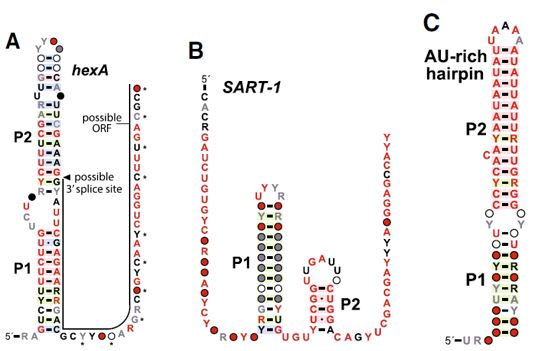
32. Beck J, Ebel F. (2013). Characterization of the major Woronin body protein HexA of the human pathogenic mold Aspergillus fumigatus. International Journal of Medical Microbiology, 303(2): 90-97. https://doi.org/10.1016/j.ijmm.2012.11.005
33. Yuan P, Jedd G, Kumaran D, Swaminathan S, Shio H, Hewitt D, Chua N-H, Swaminathan K. (2003). A HEX-1 crystal lattice required for Woronin body function in Neurospora crassa. Nature Structural & Molecular Biology, 10(4): 264-270. https://doi.org/10.1038/nsb910
34. Molinaro M, Tinoco Jr I. (1995). Use of ultra stable UNCG tetraloop hairpins to fold RNA structures: thermodynamic and spectroscopic applications. Nucleic acids research, 23(15): 3056-3063. https://doi.org/10.1093/nar/23.15.3056
35. Weinberg Z, Wang J X, Bogue J, Yang J, Corbino K, Moy R H, Breaker R R. (2010). Comparative genomics reveals 104 candidate structured RNAs from bacteria, archaea, and their metagenomes. Genome biology, 11(3): 1-17. https://doi.org/10.1186/gb-2010-11-3-r31
36. Staple D W, Butcher S E. (2005). Pseudoknots: RNA structures with diverse functions. PLoS Biol, 3(6): e213. 10.1371/journal.pbio.0030213
37. Takeda R, Petrov A I, Leontis N B, Ding B. (2011). A three-dimensional RNA motif in Potato spindle tuber viroid mediates trafficking from palisade mesophyll to spongy mesophyll in Nicotiana benthamiana. The Plant Cell, 23(1): 258-272. PMID: 21258006; PMCID: PMC3051236; https://doi.org/10.1105/tpc.110.081414
38. Adams P L, Stahley M R, Kosek A B, Wang J, Strobel S A. (2004). Crystal structure of a self-splicing group I intron with both exons. Nature, 430(6995): 45-50. https://doi.org/10.1038/nature02642
39. Ke A, Zhou K, Ding F, Cate J H, Doudna J A. (2004). A conformational switch controls hepatitis delta virus ribozyme catalysis. Nature, 429(6988): 201-205. https://doi.org/10.1038/nature02522
40. Jaeger J, Restle T, Steitz T A. (1998). The structure of HIV‐1 reverse transcriptase complexed with an RNA pseudoknot inhibitor. The EMBO journal, 17(15): 4535-4542. https://doi.org/10.1093/emboj/17.15.4535
41. Tung C-S. (1997). A computational approach to modeling nucleic acid hairpin structures. Biophysical journal, 72(2): 876-885. https://doi.org/10.1016/S0006-3495(97)78722-8
42. Wolters J. (1992). The nature of preferred hairpin structures in 16S-like rRNA variable regions. Nucleic acids research, 20(8): 1843-1850. https://doi.org/10.1093/nar/20.8.1843
43. Shu Z, Bevilacqua P C. (1999). Isolation and characterization of thermodynamically stable and unstable RNA hairpins from a triloop combinatorial library. Biochemistry, 38(46): 15369-15379. https://doi.org/10.1021/bi991774z
44. Diener J L, Moore P B. (1998). Solution structure of a substrate for the archaeal pre-tRNA splicing endonucleases: the bulge-helix-bulge motif. Molecular cell, 1(6): 883-894. https://doi.org/10.1016/S1097-2765(00)80087-8
45. Yoshinari S, Itoh T, Hallam S J, DeLong E F, Yokobori S-i, Yamagishi A, Oshima T, Kita K, Watanabe Y-i. (2006). Archaeal pre-mRNA splicing: a connection to hetero-oligomeric splicing endonuclease. Biochemical and Biophysical Research Communications, 346(3): 1024-1032. https://doi.org/10.1016/j.bbrc.2006.06.011
46. Ulyanov N B, Mujeeb A, Du Z, Tonelli M, Parslow T G, James T L. (2006). NMR structure of the full-length linear dimer of stem-loop-1 RNA in the HIV-1 dimer initiation site. Journal of biological chemistry, 281(23): 16168-16177. https://doi.org/10.1074/jbc.M601711200
47. Clever J L, Parslow T G. (1997). Mutant human immunodeficiency virus type 1 genomes with defects in RNA dimerization or encapsidation. Journal of virology, 71(5): 3407-3414. PMCID: PMC191485; PMID: 9094610
48. Cate J H, Gooding A R, Podell E, Zhou K, Golden B L, Kundrot C E, Cech T R, Doudna J A. (1996). Crystal structure of a group I ribozyme domain: principles of RNA packing. Science, 273(5282): 1678-1685. https://doi.org/10.1126/science.273.5282.1678
49. Tamura M, Holbrook S R. (2002). Sequence and structural conservation in RNA ribose zippers. Journal of molecular biology, 320(3): 455-474. https://doi.org/10.1016/S0022-2836(02)00515-6


